1.5. Medical Imaging
Medical Imaging
For much of human history, the study of internal anatomy was restricted due to cultural taboos and lack of surgical advancements. Early surgeries were dangerous, and anatomical knowledge was based more on theory than observation. The turning point came during the Renaissance, with detailed anatomical drawings by Leonardo da Vinci and Andreas Vesalius. These efforts, followed by legal reforms allowing body dissection, laid the groundwork for modern anatomical science.
The real revolution began in the late 19th century with noninvasive imaging. These techniques allowed physicians to see inside the body without surgery—transforming medical diagnostics.
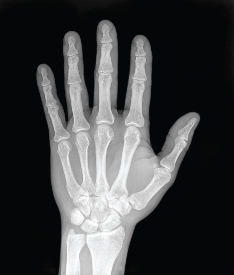
X-Rays
In 1895, Wilhelm Röntgen discovered X-rays—electromagnetic waves that can penetrate soft tissues but are blocked by dense structures like bone. The first X-ray image (his wife's hand) unveiled the potential of this technology. By 1900, X-rays became a standard diagnostic tool.
X-rays reveal bones and teeth with high contrast, making them ideal for fracture and dental imaging. Modern technology reduces radiation exposure and improves resolution, but repeated scans still carry risks.
Modern Imaging Techniques
Today’s imaging methods go beyond structure—they also reveal function and real-time changes.
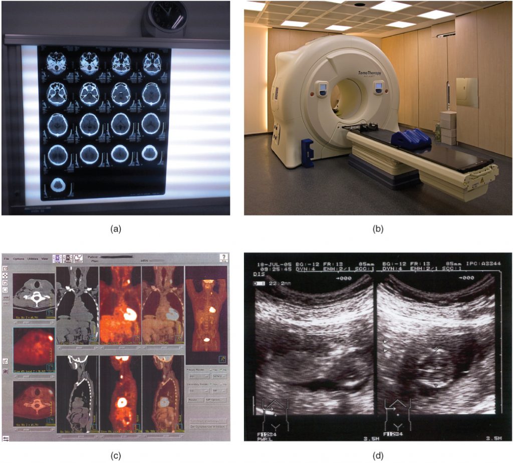
Computed Tomography (CT)
CT scans compile multiple X-ray images from different angles into cross-sectional “slices” that form detailed internal views. Useful for imaging soft tissues (like the brain or abdomen), CT scans expose patients to more radiation than standard X-rays.
Magnetic Resonance Imaging (MRI)
MRI uses magnetic fields and radio waves to map soft tissues in high detail. It is especially effective in identifying tumors and brain abnormalities. Functional MRIs (fMRI) even track blood flow to study brain activity.
Positron Emission Tomography (PET)
PET imaging uses radioactive tracers to map biological processes like metabolism and blood flow. It’s vital for detecting cancers, monitoring heart function, and assessing neurological disorders.
Ultrasonography (Ultrasound)
Ultrasound uses high-frequency sound waves to generate real-time images. It’s widely used in prenatal care and evaluating soft tissue conditions. While safe and noninvasive, it’s less effective in imaging air-filled or bony regions.
Microscopy
Though not a diagnostic imaging method, microscopy is essential in biological research. It enables the study of cells and tissues at high resolution using techniques like light microscopy, scanning electron microscopy (SEM), and transmission electron microscopy (TEM).